
What is a karyotype?
A karyotype is the complete set of chromosomes of a cell or organism, described by their number, size and shape and usually shown as a picture in which the chromosomes are arranged in ordered pairs.
By the ROQED Science team · Published · Updated
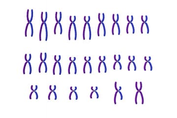
What is a karyotype?
The word karyotype is used in two ways. It means the set of chromosomes typical of a species or an individual: how many there are and what they look like. It also means the image that displays this set, with the chromosomes cut out from a microscope photo and lined up in pairs. That image is also called a karyogram.
Each species has its own karyotype. Humans have 46 chromosomes in body cells. Other species have different numbers, and the number says nothing about how complex an organism is.
A karyotype shows whole chromosomes, not genes. Each chromosome is one very long molecule of DNA wound around proteins, and it is only visible as a separate body when the cell divides.
What does a normal human karyotype look like?
A normal human body cell is diploid. It has 46 chromosomes that form 23 pairs. In each pair, one chromosome came from the mother and one from the father.
- Autosomes. Pairs 1 to 22 are the same in males and females. They are numbered roughly from the longest to the shortest.
- Sex chromosomes. The 23rd pair differs between the sexes. Females have two X chromosomes. Males have one X and one much smaller Y.
The result is written in a short formula: the total number, then the sex chromosomes.
| Karyotype | Meaning |
|---|---|
| 46,XX | Typical female |
| 46,XY | Typical male |
Sex cells are different. Eggs and sperm are haploid and carry 23 chromosomes, one from each pair, because the number is halved in meiosis.
How is a karyotype made?
Chromosomes can be told apart only when they are tightly coiled, which happens during cell division. The method therefore uses dividing cells, often white blood cells from a blood sample.
- The cells are grown in a culture so that they divide.
- A chemical is added that stops division at metaphase of mitosis, the stage when chromosomes are shortest and thickest.
- The cells are swollen in a weak salt solution, fixed and spread on a slide.
- The chromosomes are stained. The common stain gives each chromosome a pattern of light and dark bands.
- The slide is photographed through an optical microscope, and the chromosomes are sorted into pairs, today with the help of software.
At metaphase each chromosome has already been copied, so it consists of two identical chromatids joined at the centromere. This is why chromosomes in a karyotype often look like the letter X.
How are the chromosomes told apart?
Three features are used to identify each chromosome and find its partner.
- Length. Chromosome 1 is the longest. The shortest autosomes and the Y chromosome are several times shorter.
- Position of the centromere. It may be in the middle, giving two equal arms, or closer to one end, giving a short arm and a long arm.
- Banding pattern. After staining, each chromosome shows its own sequence of bands, a little like a barcode. Two homologous chromosomes have the same pattern.
The two chromosomes of a pair carry the same genes in the same order, but not always the same versions of them. That difference is the subject of the model on homozygotes and heterozygotes.
What can a karyotype show?
A karyotype reveals changes large enough to see under a light microscope.
- A wrong number of chromosomes. An extra chromosome is called a trisomy, a missing one a monosomy. These arise when chromosomes fail to separate properly in meiosis.
- Large structural changes. A big piece of a chromosome may be missing, doubled, turned around or moved to another chromosome.
- Chromosomal sex. The 23rd pair shows whether the cell is XX or XY.
| Karyotype | Condition |
|---|---|
| 47,XX,+21 or 47,XY,+21 | Down syndrome (trisomy 21) |
| 45,X | Turner syndrome |
| 47,XXY | Klinefelter syndrome |
A karyotype cannot show a change in a single gene. Conditions such as colour blindness or haemophilia, which follow X-linked inheritance, leave the karyotype looking normal.
Common mistakes about karyotypes
Several ideas get confused when students first meet this topic.
- Karyotype is not genotype. The genotype is the set of gene versions. The karyotype is only the set of chromosomes as seen under the microscope.
- Chromosomes are not always X-shaped. They look like that only after DNA has been copied and before the chromatids separate. For most of the cell cycle they are long, thin threads spread through the cell nucleus.
- 46 chromosomes, not 46 pairs. Humans have 23 pairs.
- More chromosomes does not mean a more complex organism. Some plants and animals have far more chromosomes than humans.
How to study the karyotype with the 3D model
The karyotype model is part of the ROQED Science library, a program for natural science lessons with more than 1200 interactive 3D models and courses. In Study mode you can rotate the model and look at the chromosomes from any side. In Slides mode a teacher can build slides and questions around it.
Classroom ideas:
- Ask students to count the chromosomes, then the pairs, and write the karyotype formula for what they see.
- Have them point to the sex chromosomes and explain how they decided which pair it is.
- Give a formula such as 47,XY,+21 and ask what would look different in the model.
Frequently asked questions
How many chromosomes are in a human karyotype?
A normal human karyotype has 46 chromosomes: 22 pairs of autosomes and one pair of sex chromosomes. It is written as 46,XX for females and 46,XY for males.
What is the difference between a karyotype and a karyogram?
The karyotype is the chromosome set itself, described by number and appearance. A karyogram is the picture in which the photographed chromosomes are arranged in numbered pairs. In everyday use the word karyotype covers both.
Why are cells in metaphase used for a karyotype?
At metaphase the chromosomes are at their most condensed, so they are short, thick and easy to tell apart under a light microscope. At other times they are too thin and tangled to count.
What does 46,XY mean?
It means the cell has 46 chromosomes in total and its sex chromosomes are one X and one Y. This is the typical male karyotype.